Charles E. Holman Foundation
  • Facebook
  • X
  • Youtube
  • Flickr
  • What is Morgellons?
  • Support & Resources
    • Patient Resources
    • Obtaining Social Security Benefits
    • Skin Deep The Battle Over Morgellons
    • Cindy Casey Morgellons Disease Diary
    • Press
    • Additional Morgellons Sites
  • Research
    • Research & Publications
    • For Researchers
  • About CEHF
    • Our Mission & Board of Directors
    • Advisory Panels
    • Contact Us
  • Events
    • 13th Annual Medical-Scientific Conference on Morgellons Disease
    • Previous Events
  • Patient Registry
  • Blog
  • Donate
  • Search
  • Menu Menu

Morgellons Fiber Study Summary

JENNY HAVERTY, CLINICAL MICROBIOLOGIST SCIENTIST


Date: Monday, December 13, 2004 10:32 PM

Following are microscopic observations of specimens of unknown fibers taken from four individuals suffering from a condition known as Morgellons disease.

The participants are:
(full names indicate participants who have granted full permission to publish)
Julie Karnes – who lives in San Francisco, California
Cindy G. Casey, RN – an intensive care nurse living in Sausalito, California
Murphy – an artist & musician living in Oakland, California
Wendy E. Tripp – a veterinary technician living in San Jose, California
All of these cities are in the San Francisco Bay Area in Northern California.

The descriptions are using a light microscope (Nikon LABOPHOT-2), 400x lens, and a Leitz fluorescent microscope (LABORLUX D), using an ultraviolet light source with a 330 to 380 um excitation filter and a 420 um barrier filter.

The fibers were not observed to contain septa.

Julie, collected fibers from her calves in March, 2004. I teased the fibers, and mounted them on a glass slide in sterile saline (PSS). Using the 400x lens (light microscope) the following were observed:

Red fibers 48.64 microns wide
Clear fibers 23.04 microns wide
Black fibers 28.16 microns wide
Clear slender fibers with prong like structures 7.68 microns wide
Red to red & black fibers with an internal structure that resembled
ladder-like rungs, 17.92 microns wide

Her sample also included hair, which measured from 51.2 to 74.24
microns wide. Looking at her sample of collected fibers before teasing them apart, some appeared to have a thick black speck in the center of the long strands of fibers.

I mounted the speck directly on a glass slide (I had to cut the long fibers to separate the speck from the “mass” of fibers), added saline and a coverslip, and under 400x it was comprised only of extremely tangled black fibers.

I then examined the sample described above using the fluorescent microscope (unstained in saline). The majority of the fibers were extremely bright aqua autofluorescent. The black and red fibers did not autofluoresce.

I then made a preparation of fibers in 20% KOH and Calcofluor stain (a stain used to observe fungi…yeast and hyphal elements of fungal organisms fluoresce bright apple green using a fluorescent microscope in the ultraviolet range).

The fibers did not pick up the Calcofluor stain…they were the same bright aqua autofluorescent color as observed in saline, along with the black and red fibers as observed above. I also observed some hair in this preparation, which did not fluoresce.

Cindy and her husband came to my hospital lab on August 12, 2004 and I collected a very small sample from a skin lesion on her arm. I cultured the material from the lesion using fungal culture media (Sabouroud dextrose agar, Mycosel agar, and BHI agar with blood and antibiotics), incubated at 30 degrees Celsius for four weeks. The result was negative for fungal growth, with a light amount of skin bacteria observed. There was not enough specimen for microscopic examination.

Cindy brought additional skin lesion samples for a repeat fungal culture and microscopic examination on August 31, 2004. I cultured one sample using fungal culture media. The culture was also negative for fungal growth at four weeks.

The same sample obtained on 8/31/04 was observed following teasing in saline as described above using the light microscope to have:

Clear fibers 12.8 microns to 20.48 microns wide
Blue fibers 15.36 microns wide
Using the fluorescent microscope, the fibers showed all bright aqua autofluorescent fibers with rare black non-fluorescent fibers observed.

Cindy also gave me a sample that had been collected on August 30, 2004 for microscopic observation. Using the light microscope I observed:

red fibers 15.36 to 17.92 microns wide
blue fibers 30.72 microns wide
clear tubular fibers 7.68 microns wide
clear ribbon-like fibers 15.36 microns wide
black fibers 12.8 microns wide

In addition, I found rare spore-like structures that were football shaped, 12.8 microns long, some of which had a septate-like division across the center. Also found were very rare structures slightly resembling the asymmetrical spores of Alternaria species (a fungus)…these were 48.64 microns long (Alternaria spores are 7 to 10 microns wide and 23-34 microns long). These structures were both amber colored. I also observed needle-like structures resembling crystals in this sample.

Murphy sent me prepared microscopic slides of fibers from several skin lesions. Using the light microscope observed were in summary:

blue fibers 23.04 microns wide (some of which were ribbon-like)
red fibers 12.8 microns wide
black fibers 23.04 to 30.72 microns wide
clear fibers 15.36 microns wide
large clear fibers 33.28 microns wide
Using the fluorescent microscope, each sample autofluoresced bright aqua blue, with darker fibers non-fluorescing.

Wendy sent two samples. The first sample, fibers from her torso, were collected on September 29, 2004. Using the light microscope, these showed:

black ribbon-like fibers, 25.7 microns wide
clear tubular fibers 12.8 microns wide
blue fibers 15.36 microns wide
red ribbon-like fibers 12.8 to 25.6 microns wide
brown fibers with ladder-like rungs 12.8 microns wide
brown fibers with prong-like structures along the sides 3.84 to 10.24 microns wide

The above sample had a predominance of the black fibers described above.

A second sample (also from torso) collected on October 7, 2004 showed the same types of fibers as described above with the addition of:

brown fibers with ladder-like rungs 33.28 microns wide

Using the fluorescent microscope, observed were bright aqua autofluorescent fibers, with black and red non-fluorescing fibers.

In summary:

There were many similarities in fibers from all four individuals, both in size and color. All samples showed bright aqua autofluorescence using the fluorescent microscope, with red and black non fluorescent fibers. The fibers collected from these four individuals from different counties of the San Francisco Bay Area are so similar to each other that the causative agent may be epidemiologically the same.

Respectively submitted,

Jenny Haverty
Clinical Microbiologist Scientist
Marin General Hospital
Greenbrae, California

/wp-content/uploads/2020/09/CEHFLogo.png 0 0 Charles E. Holman Foundation /wp-content/uploads/2020/09/CEHFLogo.png Charles E. Holman Foundation2021-01-19 16:14:142021-01-19 16:14:39Morgellons Fiber Study Summary

Morgellons Research Update September 2005

SEPTEMBER 9, 2005

A few very preliminary results and observations on Morgellons research:

The goal of this research is to determine the cause of Morgellons Disease. At this point we do not know whether there are environmental or chemical toxins, allergens, or infectious agents involved, or even a combination of more than one of those. We are not pre-biasing the studies by making assumptions about the causative agent(s). The chemical analyses are looking for environmental contaminants, anything from heavy metals to organic chemicals. A possible role for infectious agents includes considering the possibility of infection by: insects, bacterial, fungi, nematode-like worms, parasites and viruses.

There are two widely discussed possible causes that are discussed on the internet extensively. These include an infection of the bacterium Stenotrophomonas maltophilia.and an infestation of the arthropod Collembola. Since these have been so widely discussed, they seemed likely logical starting points for part of the research and will be discussed below.

  • Two primary types of bacteria have been cultured from skins samples of multiple Morgellons patients. The bacteria are of two types; a chain or 2-4 rod-shaped bacilli and tiny, spherical, cocci/diplococci. On solid media the cocci make a hard membrane-like coating over the colony. The liquid culture of bacilli usually contains a very stringy material after a few days of culturing. Some macroscopic fibers have appeared in these cultures, but it is unclear where they are coming from. We are trying to determine whether they are environmental contaminants or a product of the bacteria. It is also possible that the long fibers are nothing more than DNA from the dead bacteria. We are currently performing PCR (to amplify the microbial DNA) and DNA sequence analysis of both of these isolated bacteria. These results should be available by the end of September or early October.
  • As mentioned above, Stenotrophomonas maltophilia.has been mentioned as a candidate, causative bacterium in Morgellons Disease. Amplification of DNA from the bacterial population isolated from the skin, scabs and shed material of the Morgellons patients and sequencing is still incomplete, although it will hopefully be completed during the next month or so. Bacterial isolates cultured from skin samples from four Morgellons patients residing in climatologically and geographically distinct areas of the United States have revealed no evidence of S. maltophilia thus far. Based on published reports, when S. maltophilia is cultured on blood agar plates there is a distinct flagellum (or multiple flagella) that is visible on the rod-shaped bacteria (bacilli). The bacilli that we have observed do not appear to be flagellated and are much longer than we would have expected for the characterized and published strains of S. maltophilia. They are also clustered in ways that do not look like the published images of S. maltophilia. DNA sequencing will give provide an answer as to whether this bacillus is present in Morgellons patients or not.
  • The putative role of the arthropod Collembola in Morgellons disease. The paper by Altschuler, et al., (J. New York Entomol. Soc. 112(1):87–95, 2004) entitled, COLLEMBOLA (SPRINGTAILS) (ARTHROPODA: HEXAPODA: ENTOGNATHA) FOUND IN SCRAPINGS FROM INDIVIDUALS DIAGNOSED WITH DELUSORY PARASITOSIS, described evidence of Collembola in patients with symptoms resembling Morgellons disease. We decided to look at the molecular level for DNA evidence of Collembola in samples of skin, scabs and other shed material from Morgellons patients. To do this, PCR primers were synthesized that would amplify DNA from the cytochrome oxidase II gene from any of the 20 families and over 1,000 species of Collembola (Frati, et al., Evolution of the mitochondrial cytochrome oxidase II gene in Collembola, J. Mol. Evol., 1997). The PCR primers are designed as follows, as per the Frati, et al. paper:

AATATGGCAGATTAGTGCA
GTTTAAGAGACCAGTACTT

When used in the PCR reaction, these primers are expected to amplify a 690 base pair fragment of DNA. Thus far, no DNA fragment has been amplified from the Morgellons samples. This suggests one of two likely scenarios:

1) that the samples contain no Collembola DNA for this gene, or

2) that the PCR is not working properly. To examine the 2nd possibility, PCR from a sample of old leaves, and a bit of soil has yielded an (approximately) 700 bp fragment of DNA as expected. This fragment of DNA will be sequenced in the near future. If it is a Collembolan species, then the PCR is working and it suggests that the samples isolated from Morgellons patients, thus far, do not contain Collembola DNA, and hence, these Morgellons patients symptoms are not from Collembola. More unrelated tissue samples from other patients will be assayed for Collembola DNA in the future.

There are 4 other areas of research that are ongoing or will be carried out during the upcoming months, and being carried out at laboratories in Oklahoma, California and Italy. These are 1) a more detailed spectroscopic and microscopic analysis of the Morgellons fibers, 2) assaying of material from Morgellons patients for modified amino acid neurotoxins, 3) chemical analysis of the Morgellons fibers to determine the chemical composition and presence of unusual organic compounds and heavy metals and 4) analysis of tissue samples at the nano-molecular level to screen for foreign material that could be involved in Morgellons disease.

This research briefing is not meant to be overly interpreted or viewed in any way as conclusive. It is an interim report on the status of the research and nothing more. When there is firm, incontrovertible data, it will be presented as such. For now, this is to let Morgellons patients, family, friends and health-care providers know that there is organized, systematic research being performed and where the research stands.

All of this research is being conducted through the efforts of the Morgellons Research Foundation, with no funding support from either the federal government or any pharmaceutical or biotechnology corporations. None of the researchers associated with the Morgellons Research Foundation have received any salary from the Foundation. All of the research to date has been performed as a community service and a desire to help the sufferers of Morgellons Disease. The experiments needed to systematically research this disease are both time-consuming and expensive. The seemingly slow progress of the research is not by intent or inattentiveness; it is due to the time-consuming nature of the experiments themselves, and limited availability of research personnel and funding. The goal of this research is not mere intellectual curiosity. The goal is to identify (conclusively) the cause(s) of Morgellons Disease. Once the causative agent(s) are identified, therapies can be devised to treat and ultimately cure Morgellons Disease.

Randy S. Wymore, Ph.D.
Assistant Professor of Pharmacology & Physiology
Oklahoma State University
Center for Health Sciences and
College of Osteopathic Medicine
1111 W. 17th St.
Tulsa, OK 74107

/wp-content/uploads/2020/09/CEHFLogo.png 0 0 Charles E. Holman Foundation /wp-content/uploads/2020/09/CEHFLogo.png Charles E. Holman Foundation2021-01-19 16:09:422021-01-19 16:09:42Morgellons Research Update September 2005

Morgellons Research Update April 2006

APRIL 5, 2006

Recently, I have been asked to evaluate some research that may have bearing on Morgellons Disease and that is presented in an online streaming video. Before I give my comments, I would like to state that I am NOT calling into question either the integrity or honesty of the person(s) who made the video.

I have viewed the video and am not able to come to any conclusions about the video for the following reasons:

1). When I am asked to review manuscripts for publication or grant proposals, there is always a clear statement of hypothesis to base the data on. Biomedical research is generally hypothesis-driven and the research is aimed at testing the hypothesis. I could not ascertain what hypothesis is being tested in the video.

2). The microscopy images are of a high quality. However, when such data is presented at a meeting, or in a publication, a scale-bar would always be included. This may sound trivial, but I assure you that it is absolutely critical to know whether the objects are viewed with minimal magnification or at 1,000X oil immersion. There are times when my guess is that some objects are 60-100X magnification and other images seem highly magnified at probably 400-1,000X. Seeing microscopic images without knowing the size is like looking at a road-map of a new land without a mileage scale marker. One just doesn’t know if it is 400 meters from point A to point B or 100 miles.

3). Whether a PowerPoint presentation or a paper, each figure displayed will have a thorough description of what is being shown. Species, tissue, cell culture, temperature, culture media, etc. are all details that would be written out so that the image has some meaning. This is true whether it is a chart or photo or a micrograph. In the absence of such information, one is left to try to guess what is being shown. This lack of information or interpretability is true from the start to finish of this video, other than occasionally showing a man in a laboratory. There are objects in some liquid that are shown. Is the liquid, water, saline solution, tissue culture media or bacterial media? Are the objects that may be cells from a cell culture or taken from a living organism? It can NEVER be left to the reader or viewer of a scientific presentation to have to GUESS or SPECULATE about what is being viewed. It must be clearly spelled out. This is true in all branches of biomedical research, including: molecular biology, microbiology, proteomics, genomics, pharmacology, etc.

4). There are words and phrases that show up in the video that have no meaning and hence, come across as though they have meaning, but as presented they simply do not. They should have been explained and there is no attempt to do so. For instance: a. “A model organism based on quorum sensing”. This phrase is missing the end of the sentence. “…quorum sensing”…. ‘something’. What is the ‘something’? Some bacteria utilize quorum sensing. Quorum-sensing is one way that bacteria control the explosive growth phase (log phase) with what is called ‘stationary phase’. It is how they keep from overpopulating an environment. What is the so-called model organism quorum sensing? Sugars, protein, heat, acid, nitrogen or a specific molecule that the organism releases to be sensed by others of its own kind? Bacteria might release molecules into their environment to guide how the population, as a group, will behave. The gut bacteria, E. coli use quorum sensing. The phrase “… based on quorum sensing…” has no intrinsic meaning and needs to be explained. b. “Involved technology represents proteomic and synthetic genomic research.” Again, these words are meaningless without explanation. Proteomic and genomic research goes on in hundreds of labs around the world. Both of those disciplines have meaning only when the specific ‘genome’ or ‘proteome’ is defined. What species or multiple species are involved in this research. What does synthetic genomic research mean? The word ‘genomic’ implies a genome. A genome is the entire DNA pool of genes of a specific organism. There is no such thing as a synthetic genome. There may be SPECIFIC individual genes that are engineered for use from one species to another species, but those genes are still part of the human or mouse or Lepidoptera or whatever species is being discussed. c. “Next footage will demonstrate how single cell, quorum sensing microorganisms grow and replicate a multicellular entity in the human body”. Does this imply that the footage was taken in a human body? It did not appear that this was the case. The problems described earlier arise again. Is this tissue, in a human, in another animal, or if they are tissue culture cells, are they human, rat, mouse, or what? The phrase “…single cell, quorum sensing microorganisms grow and replicate a multicellular entity…” just has no meaning. We need to know what is going on here in detail, or it is not informative. d. “multinucleated giant cell” I watched this section several times & could not see any multi-nucleated giant cell. Perhaps still image, micrographs would indicate this cell, but I could not see it in the video. There are multi-nucleated cells in various tissues from many species. Such cells with multiple nuclei and intracellular organelles are usually quite easy to observe, but I could not make out any organelles or nuclei in the video. e. “quorum sensing robotic like behavior”. This is a statement that may well cross scientific disciplines and may be out of the realm of my expertise. On the other hand, robotic devices cannot exhibit quorum sensing, so I was not surprised when I inquired from a robotics mechanical engineer about the phrase & she had never heard of quorum sensing. Again, quorum sensing is the ability to communicate amongst a population by sensing certain molecules. In some bacteria it is possible to knock-out the ability to utilize the natural quorum sensing abilities of a species and often this will result in runaway cell overgrowth leading to death of most of the population.

5). While never coming out and discussing the specifics, it seems that the implication is that an ‘organism’ has been generated by someone with genes of different species. If so, then there is no need to even debate the matter with nice production videos, still micrographs or speculation. DNA sequencing is a common-place procedure in 2006. There are thousands of labs and numerous commercial facilities that will sequence DNA at a fairly inexpensive cost (under $20.00 per reaction). If there is an engineered organism that the maker of this video has in his possession, then it would be trivial to sequence across the DNA splice sites. If sections of DNA from 2 species have been joined together, then the sequence should reveal the first organism to one point and then the other organism beyond that splice site. DNA technology lends itself to accidental splicing of mismatched pieces of DNA fairly frequently. Labs that do DNA and RNA research are frequently checking to make sure that such accidents are detected, or much valuable research time can be wasted. Intentional and accidental DNA splicing occurs in the modern molecular biology world & simple DNA sequencing reveals when such errors occur. Since fairly poor labs and even college teaching labs can sequence large stretches of DNA, there should be no reason that the maker of the video cannot do so. Such work is inexpensive and easy. It is also CONVINCING and NOT subject to interpretation. Whenever an image or video has to be interpreted, there is the possibility of misinterpretation. DNA sequencing either works or it does not work. A fragment of sequence can be submitted to the global database search engines and will be either human, or mouse or rat or cockroach or whatever species it came from. There is no subjective interpretation necessary. There was a time, well over a decade-and-a-half ago, when DNA sequencing was difficult, time-consuming and expensive. Now it is cheap and accurate. There is no reason not to use the ‘gold-standard’ of DNA sequencing to identify this supposed, engineered organism. Until the accepted molecular tools are utilized to give an objective result, the maker of the video will be left with subjective conclusions that can be interpreted in different ways. Sequencing DNA on both strands, from both directions gives a result that cannot be argued. Looking at microscopic images and video can never give such incontrovertible proof of something. Properly documented and well-described mages and video can be a PART of a case, but will never be considered proof. From the 1970s and back into earlier decades, molecular tools did not exist. Now that they do, it is incumbent upon any researcher to make use of the standard, commonplace tools for a specific type of research. Availability of the organism’s DNA or RNA should not be a problem. When properly handled, the DNA and/or RNA from just one single cell should be enough to amplify and then sequence. The video footage under discussion is fairly extensive and claims to show a novel organism, therefore, getting one single cell for DNA sequencing does not seem like it should be too difficult.

Whenever I or any other scientists publish our work, it is up to us to convincingly demonstrate that we have accomplished what we say we have. I could never publish something and leave it up to the reader to interpret what I am trying to say. Such research would never get through scientific review and could never be published. No, all relevant facts must be clearly described and defined. Most of my publications have been in the fields of molecular biology, physiology and pharmacology. Any competent scientist should be able to follow the protocol steps described in my papers and replicate my experiments. If they could not, it would call into question the validity of the paper. Results must be replicable and all steps should be clearly described for other to repeat the experiments. Hopefully, if the maker of the video feels that he has information regarding the cause of Morgellons Disease, then he will perform the necessary, proper and fairly easy experiments so that this information can be shared with other researchers. Just as with many things in life, talk is cheap. When it comes to scientific hypotheses, talk is more than cheap, it is meaningless. One must not only ‘talk the talk, but must also walk the walk’.

These matters are of vital importance as it will likely be necessary to identify the cause of Morgellons Disease before effective treatments can be developed.

Best wishes for success to everyone researching the cause and treatment of Morgellons Disease.

Sincerely,

Randy S. Wymore, Ph.D.
Assistant Professor of Pharmacology & Physiology
Oklahoma State University
Center for Health Sciences and
College of Osteopathic Medicine
1111 W. 17th St.
Tulsa, OK 74107

/wp-content/uploads/2020/09/CEHFLogo.png 0 0 Charles E. Holman Foundation /wp-content/uploads/2020/09/CEHFLogo.png Charles E. Holman Foundation2021-01-19 16:07:442021-01-19 16:07:44Morgellons Research Update April 2006

Morgellons Research Update April 2004

APRIL 17, 2006

From: Randy S. Wymore This is an open letter to all interested parties regarding Morgellons Disease and my role in Morgellons Disease research. This may come off as a long and rambling letter, but a number of emails to me, as well as comments about me, suggest that I need to cover several issuess.

I was sent a copy of statements and questions that a poster by the name of Sabrina made on a public forum. Other email writers asked individual questions or made comments in bits and pieces, but Sabrina pretty much covered all of the topics, so I will reply to her statements and questions directly.

First of all, Sabrina, thank you for pointing out to me some of the issues that I had not even thought about conveying. You asked questions that I should have answered, but in some cases never even thought of in the first place. I am pasting in some of your comments and questions and then will add my thoughts after each one.

“Since fairly poor labs and even college teaching labs can sequence large stretches of DNA, there should be no reason that the maker of the video cannot do so. Such work is inexpensive and easy. It is also CONVINCING and NOT subject to interpretation.”

‘This tells me that MRF should have “no reason” to not already have this information as well, right? Dr Wymore says it “is inexpensive and EASY.” I do not really know, perhaps I missed this release of information. Does anyone else know? ‘

No, you did not miss the release of this information. I should have made clearer what I meant. I do not have a candidate organism in hand that I can use to extract DNA to amplify and sequence. The red & blue fibers, black specks and white granules are associated with the disease, but none of those objects are ‘it’, the actual organism.

The maker of the video claims to have ‘it’ in hand. Once a researcher has the suspected organism, then one can do the molecular biology. This is actually the key to this whole mystery. What is the causative organism? This is where my research and the video maker’s are quite different. He claims to have isolated the causative agent. I have not done so. Or, if I have, I have not recognized it as such yet. Also, several people have questioned my ‘sequencing of DNA for less than $20.00’ comment. Well, here is a commercial service at: http://www.nwdna.com/sequencingpricing.htm . The type of sequencing that would be performed is actually listed at $8.50 per sample. I overestimated the cost because there is also 1-4 day mailing of the samples to work into the total price.

Also, once again I did not really make my comments very clearly. I did not mean that the entire organism’s genome of DNA could be sequenced for that small price. But, for that price one can get several hundred, to a thousand nucleotides of DNA sequenced. Definitely enough sequence to compare to the global databases to get a handle on an organism. The entire genome would take big bucks and months to years to finish, depending on the size of the organism’s genome.

‘”A fragment of sequence can be submitted to the global database search engines and will be either human, or mouse or rat or cockroach or whatever species it came from.”

So, what has the Morgellons fragment been determined to be??? It should be pretty easy and inexpensive to get this information, right? Perhaps I missed this information as well. ‘ I really do hope that it will be easy and inexpensive to get this information once a candidate Morgellons organism is identified. ‘

“There was a time, well over a decade-and-a-half ago, when DNA sequencing was difficult, time-consuming and expensive. Now it is cheap and accurate. There is no reason not to use the ‘gold-standard’ of DNA sequencing to identify this supposed, engineered organism.”

‘ I would love to ask the good doctor about this cheap and accurate technology he speaks of. I see “no reason” why this has not already been done. Do you? Maybe it has? Who knows? We don’t. ‘

You bring up a point that, in all honesty, I never even thought of until I read the above statement. You don’t know what I have done or not done. This is an issue that I should have thought about but had not. Please stay with me for a bit while I try to explain why I never thought to discuss my lack of progress with the Morgellons community.
A) I am about to use the term ‘professional’ with regard to biomedical research and I just want to make clear what I mean. I mean ‘professional’ in the basic sense of ‘the way one makes a livelihood’. I don’t mean professional in the sense of a doctor or lawyer; that sort of thing. I am paid to be a scientist and in general, I have resources that most amateur researchers do not have at their disposal. Now, also I am not using the word amateur in a negative sense, the way some might say ‘amateurish’. The basic meaning of amateur is that a person does not do the work or endeavor for a living; it is not the source of their livelihood. My use of professional vs. amateur research is not meant to disparage anyone’s efforts in science. Many of the asteroids and comets that have been discovered in recent years have been done so by amateur astronomers. Some of them have better equipment than many academic institutions, but they are still amateur in that they do not get paid to be an astronomer.

B) There is an aspect to being a professional scientist that many people are unaware of. We don’t publish our failures. For the most part, moving the cutting edge in any field forward is dependent upon what does work & not the failures. Silly as this might sound, I really never thought about updating on the lack of progress as I would never do that in my field. This was pretty clueless on my part as I know that many Morgellons sufferers, their friends, family members and health care providers want to know what is going on.

If I was doing this purely from an academic perspective I would research this for months to years & never write or publish anything until a complete enough package of information was accumulated that I could write a manuscript & send it out for review & publication. As an example, my last co-authored publication
(http://www.molbiolcell.org/cgi/content/full/16/6/2972) took a couple of years and 15 co-scientists in Florence, Turin, Milan, Italy and Tulsa, USA to accomplish the work.

The manuscript was first submitted in October of 2004 and after more experiments and revisions, was finally published in the June of 2005 issue of the Journal. I chose this paper as an example, not because it has anything to do with Morgellons, but because it is freely available at the above link. Many journals have subscription-only access to articles that they publish, but I think this one can be accessed from anywhere.

We never discussed this research with any outside groups until the paper was published. This slow pace is typical for professional scientists. The general feeling is that it needs to be as perfect and complete as possible before it is ready for public consumption (and here ‘public’ means other scientist). I have NEVER planned secrecy regarding successes with my Morgellons research.

From the beginning, I have planned to release important information as soon as I am sure about it. I had NOT planned to make everyone wait for the glacial pace of writing a manuscript, getting it reviewed and then published; which can take a year or more. Since I have not identified the causative agent(s) of Morgellons, I didn’t think I had anything to report.

I see now that reporting failures or limited success should be done. In my field there are no forums or posting boards or anything like that, so I had not thought about keeping those with personal interest in Morgellons informed in a ‘real-time’ way. In my mind, the science and my work on Morgellons were separate from the conversations surrounding Morgellons and that are going on around the clock and around the world. I realize now that I was wrong about that.

A bit further below I will describe the current state of my research, including the dead-ends and failures that I would never normally discuss with the scientific community; not because I am embarrassed or feel like a personal failure, but because it would be considered irrelevant and not of interest within the scientific community.’

“Availability of the organism’s DNA or RNA should not be a problem. When properly handled, the DNA and/or RNA from just one single cell should be enough to amplify and then sequence.”

If this, “should not be a problem” what is the answer? Does the good doctor have one? Who can answer this????”? ‘

did not explain what I meant and your question is quite appropriate. What I meant is that if the maker of the video actually HAS in possession a few cells of the cause of Morgellons, or even better a culture of the organisms, then he should have plenty of DNA (or RNA if ‘it’ uses RNA instead of DNA) to do what I described.

The DNA will need to be amplified by a process called PCR (polymerase chain reaction). All it takes is a few cells worth of DNA or RNA to be able to amplify a stretch of DNA for sequencing. I might have misinterpreted the video, but the way it was presented it left me thinking that he has the organism isolated or at least he has access to the organism. IF that were the case, then it should be no problem to extract the DNA/RNA perform PCR and sequence it. Also, when I say ‘not a problem’ I mean it within the context of lab work & not the way I would use the phrase outside of the lab.

I shouldn’t have done that. Molecular biology can be very tedious and repetitive. I might take weeks to get a single PCR reaction to work so that enough DNA can be generated to sequence. In my glacially-paced world, generating the sequence, whether in 3 days or 3 months would be ‘not a problem’. Bad phrase for me to have used. However, this does NOT change the meaning I intended.

IF the maker of the video DOES have in his possession, or access to the organism that causes Morgellons, then there should be plenty of genetic material to amplify and sequence.

‘ “Hopefully, if the maker of the video feels that he has information regarding the cause of Morgellons Disease, then he will perform the necessary, proper and fairly easy experiments so that this information can be shared with other researchers. Just as with many things in life, talk is cheap.” ‘

‘ Well I’ll be. Who knew that these experiments are “easily” done? We wouldn’t have known this before. I thought this was going to be hard, what do I know? Thank God this is so easy for them. Should speed things up do you not agree????”

Within the context of molecular biology, PCR and sequencing IS considered easy. It can be frustrating and time consuming, but ‘easy’ in the sense that it is ultimately doable. In my previous lab, one of the technicians had a degree in English, but ended up doing lab
research. She never had a formal molecular biology course, but after a few hours of training she could PCR with the best of them. Many aspects of molecular biology are considered ‘cookbook’ with regards to the techniques. PCR and preparation for sequencing fit into that category.

The difficult part is identifying the causative agent. He claims to have already done that hard part. The DNA analysis in comparison, is the easy part.

Sharing information with other researchers????? Why in the world would tamtam even be bothered with suffers to begin with? None of us are researching anything, right? Could the good doctor be out of touch?

There was no intent on my part to disparage the research that anyone has done or will do in trying to figure out Morgellons Disease. I see that I did not specify, but I was referring to the research community of scientists who do research for a living. Quite frankly, I am in awe of some of the wonderful, high resolution micrographs I have seen from some Morgellons sufferers (or their loved ones). Many of those images are far better than any microscopic images I have taken. I do NOT say that in a sarcastic or patronizing tone. I mean it. I am not a microscopist by trade & will never be as good with a microscope as some of you who may be reading this. The information I meant was the information about the only thing that really matters; WHAT IS THE CAUSE OF MORGELLONS DISEASE? He claims to be in possession of that knowledge. If I even thought that there was a strong MAYBE that I had found the cause of Morgellons I would get that information out so that treatment options could be considered. There will be plenty of health-care providers willing to take a chance on a treatment, once the cause has been identified. Then, at a more leisurely pace I’d finish up all proper details for the academic community.

‘Perhaps he is only intrigued by the actual organism. ‘

In this instance, ‘he’ means me. A fair enough issue to bring up, since most readers of this long-winded essay and I have never met. I will give an answer, but each person has the obligation to choose to believe me or believe that I am not telling the truth. I am definitely NOT ONLY interested in the actual organism itself. Personally, I wish that ‘it’ did not exist. I wish that no one suffered from Morgellons Disease. I clearly have no power to grant wishes.

‘Talk may be cheep but in this case silence is not golden either. This silence creates a negative space that can be filled with all sorts of things and this is dangerous in my opinion. ‘

Absolutely true! I am trying to fill-in some of that void.

‘MRF was created for Morgellons sufferers. It is a tax free organization and belongs to all of us. Do you feel like a part of this organization? People repeatedly ask to help them in any way they can. Are these offers ever accepted? Not that I am aware of. Maybe they have all the help they need? I truly hope this is the case. ???” ‘

Others will have to answer most of those questions you ask. The one question that applies to me is whether or not I feel like I am part of this organization. Since late last spring I have been the Foundation’s Director of Research in a voluntary capacity, so yes I am part of the MRF. I am not a board member or responsible for the day-to-day running of the Foundation, so in that sense, no, I am not a part of the formal structure of the Foundation. I could choose to no longer volunteer or they could ask me to stop being the Director of Research and that would be the end of it.

‘ Apparently we walk a different path. Now be on your way. ‘

I think that some of our paths are not to different and overlap on at least one topic. We both want to know how to cure Morgellons Disease.

‘I would have much rather had Dr. Wymore tell us if the DNA sequencing has already been done instead of only mentioning this to disprove a video. Does anyone else find this to be strange and sad?’ ‘I see contradictions everywhere! It’s easy, inexpensive, and accurate…… Tell me why we are all still sitting here in the dark suffering and grasping at straws daily? ???” ‘ ‘Why did I come to a public forum to look for answers rather than a foundation set up for people like me? Think about that one very hard.???????’

While one or two of your questions are likely rhetorical, I will address
the others. Here is my research update that includes failures,
dead-ends, unknowns and observations that will never be published, because no one in the research world would be interested. Some of what I am about to tell you has only been done once and until it is replicated is fairly meaningless in scientific circles. Yes, as I will discuss below, there has been DNA sequencing. No, it has not revealed the likely cause of Morgellons.

1) Individual red and blue Morgellons fibers were placed in bacterial media and cultured at body temperature. Isolates of those bacterial populations were grown on lab preparative media, blood agar, chocolate agar and a type of media that tends to support fungi better than bacteria. The bacteria were stained with various stains and observed both alive and dead. The bacteria were separated out into pure cultures (I think). PCR was performed and the amplified DNA was sent to a commercial sequencing lab to do the DNA sequencing. Two different
bacterial species were identified. They were: a) Pseudomonas putida and b) Corynebacterium efficiens. Does this identification mean anything? I do not know. Both of these can cause infections, especially in immunocompromised individuals. Both of these bacterial types are found in soil and can be found in skin.

The fact that they grew from fibers that were associated with skin makes it difficult to say if they are related to Morgellons Disease in any meaningful way, or merely normal skin contaminants.

Still, these are the only two DNA sequences to be identified so far.

2) The fibers can be dissolved in 6 molar hydrochloric acid, at 90 C. The fibers first fragment and then dissolve, much like hair.
The fibers are not touched by strong reducing agents or chemicals that are routinely used to isolate DNA (guanidinium isothyocyanate), even after sitting in the chemical for over a week. One preliminary spectroscopic analysis suggested a strong sulfur signal.

This has not yet been reproduced. A blue fiber was analyzed a little over a week ago by a collaborative chemistry lab.
This fiber was neither, cotton, cellulose nor any known textile. No definitive composition has been determined so
far. The chemical/spectroscopic analysis is an ongoing process.

3) I receive 2-15 packages of samples per week. We look at the samples in the order they come in but each sample may have 5-50 smaller packages of samples inside them.

We cannot examine all of them in detail. One emailer asked if my lab was poor and in a basement somewhere and another
person commented that I have been given all of the material I need to solve the mystery, but it is up to me to do so, I just need to hurry up and do some work.

Just to be clear, no, my lab is not in a basement. As for the poor vs. rich, that is a tough call. Rich labs have million dollar grants, and I do not have one of those, so maybe poor would be a better description of my labs financial status.
On the other hand, I amnot lacking for supplies and normal operational needs.
My lab personnel consist of my full-time lab technician, an undergraduate who volunteers a few hours a week and myself.
We are a small lab for sure. The small number of hands in my lab that are working on Morgellons is a factor in how much gets accomplished.
If we quickly examine the submitted samples and move on there is the risk of overlooking something important.
If we spend a lot of time on individual samples we keep getting the backlog of samples even further behind.
Trying to strike a well-balanced approach is not trivial and the time spent per sample is variable, depending on the nature of the sample.
I can only ask that people who have submitted samples be patient.

4) There are many ideas about the cause of Morgellons that have been forwarded to me.
Some of the proposals that people have sent me include:

a) springtails,
b) S. maltophilia,
c) Strongyloides, cutaneous larva migrans and nematodes in general
d) flukes,
e) parasitic nematomorphs from cotton,
f) engineered organisms (either industrial-strength that got into the environment or intentional bio-weapons),
g) extra-terrestrial,
h) ancient life-forms that escaped from an earthquake fault or volcano,
i) viral,
j) ‘other’ protozoans or parasites,
k) non-contagious genetic or environmental factors and l) dangerous dental work.

The bad news is that each of those ideas has proponents who feel they have proof for their idea. The good news is that the more specific and refined the idea, the easier to get evidence for or against the likelihood that it CAUSES Morgellons Disease.

There is a distinction here, which needs to be perfectly clear. If a person has non-healing lesions, is in a weakened state or has a compromised immune system, there are many organisms and parasites that may set up house in/on that person. I am not interested in those opportunistic organisms
.
I want to find the CAUSE of the diverse and often strange
symptoms of Morgellons Disease. The cause must explain all of the
varied symptoms including the production or appearance of the red, blue, clear and dark fibers, the black specks, the sand-like granules, the callous-like membrane, the peripheral neuropathy and the central nervous system changes.

When I am trying to PCR and then sequence DNA I must use a small stretch of known (or suspected) DNA called a primer. Primers can be very general or very specific. When we think something is bacterial, there are primers that can amplify and then be used for sequencing the DNA that will work on ANY bacteria known. These same primers are used to find new and previously unknown bacterial types.

The reason is that all bacteria, from the ones living in everyone’s small intestines to the exotic ones found in very novel locations, all share certain genes and stretches of DNA. Such primers are perfect for searching for unknowns that we grow in the laboratory from the fibers or scabs of Morgellons sufferers. But, if we think it is a specific organism, the primers can be designed that will ONLY amplify that specific DNA.

With that in mind, I feel that a) & b), springtails and S. maltophilia can be eliminated. The primers for the stretch of sprintail DNA will amplify a specific gene from over a thousand species of Collembola and yet we can never amplify this DNA from fibers, scabs, dried skin or callous material.

Even if the Collembola had been in contact with the scabs or the fibers it is likely they would have shed a few cells and that should be enough to amplify the Collembola DNA. Since that hasn’t happened, I consider it unlikely that Collembola are the cause of most of the Morgellons symptoms. Also, none of the Collembola proponents have explained to me where the fibers are coming from or why the neurological effects would make any sense from a Collembola infestation.

Similarly, in addition to the general bacterial PCR primers, we have S. maltophilia-specific primers and cannot amplify any DNA. I also do not think that the proponents of allergies to bad dental
adhesives/antibiotics have made a very strong case.

First, the sulfa drugs need to be at a high enough concentration to cause an allergic reaction & as a pharmacologist I find it hard to believe that such a concentration could ever be reached from sulfa-drugs leeching out over years and decades from the dental work. Second, some patients actually report some improvements while on sulfa-style antibiotics. Well, it can’t work in both directions; a person either is or is not allergic to sulfas, and the drugs will either cause the problem or help it.

Third, there are children who have never had any dental adhesives/ crowns/ caps on their teeth that have symptoms of Morgellons Disease.
Remember, I’m not trying to convince anyone of anything here. I have been asked to not be secretive and say some of what is on my mind. That is what I am doing here. This is NOT a position statement from the Morgellons Research Foundation. These are my personal thoughts and nothing more. I am trying to give you a glimpse of my thought process as a scientist.

I will address the Strongyloides and CLM proposals in a separate section. Most of the other proposed Morgellons causative agents are not easily testable, until a specific species is identified or suggested.
D-k in the above list all remain candidates. I’m not saying any specific ones are LIKELY, but from a scientific perspective, until they are eliminated (which won’t be easy for some of them) they remain candidates. There is also the possibility that a combination of the above may be necessary. There may be a genetic predisposition or a genetic protective component to Morgellons. Similarly, there may be certain environmental factors that trigger or repress the symptoms.

5) Some of you may have had the misfortune of receiving one or more vile, vulgar and belligerent emails from a person or two who claims to have figured out what causes Morgellons Disease and who is also kind enough to share that information, but only for the right price. The website in question claims that there really is no such disease as Morgellons and that it is nothing more than a combination of common parasites and nematodes. Specifically, Strongyloides is mentioned alongwith cutaneous larva migrans.
I would like to quote from this website (as of 4/12/06): “By the way, fibers are the shedding of their shell/ skeleton!” That is utter nonsense for multiple reasons. Nematodes/small worms/Strongyloides have neither a shell nor an internal skeleton or an exoskeleton. They have a multi-layered cuticle. Even if one wanted to make a point over semantics to give the benefit of the doubt to him/them it still makes no sense.
The Morgellons fibers cover a range in size from way tinier than even the smallest Strongyloides to several inches in length. The Morgellons fibers are not hollow, nor do they look like a split open cuticle. The red and blue fibers have no cellular structure or even ‘ghost’ outlines where the cells might have been. Many of you have probably spent more time looking in a microscope than I have. Most Morgellons sufferers that have looked at the fibers feel the term ‘fiber’ best describes their appearance. Small or large, red or blue they look like fibers. Also, published data suggests that Ivermectin will cure 97% of Strongyloides infections with a 2 day course of treatment. Some Morgellons sufferers have taken Ivermectin for weeks or months and few have reported that they are cured afterwards. There has been no DNA that can be amplified from the fibers that has been anything other than normal contaminating bacteria or in one case human female DNA, likely from cells of the person who submitted the fibers (it looked like there was a bit of bleeding & some white blood cells may have been the source of the human DNA). I do not know if the fibers are synthesized, shed or how they form, but they are not the shell/skeleton (which is impossible) or cuticle of Strongyloides. In an effort to be thorough, I will not completely reject the hypothesis that Strongyloides are involved in some cases of Morgellons.
I do, however, reject the idea that they are the CAUSE of Morgellons Disease. I will order PCR primers for Strongyloides and PCR from the scabs and fibers to see if there is any evidence of cutaneous larva migrans.

6) Some Morgellons patients have been fortunate enough to find kind and competent health-care providers. This is wonderful if it has been your experience. Unfortunately, many Morgellons Disease sufferers have not been treated in such a kind manner and have often been labeled as
delusional, without the benefit of a through physical examination. Many of you have been told, ‘stop scratching and you will heal’.
Well, while my ultimate goal is to find a cause and cure for Morgellons Disease, one of my short-term goals has been to convince a skeptical medical world that Morgellons Disease is not an internet-based subset of Delusions of Parasites.

Over the last 6-9 months, I have been accumulating data (really evidence) that SOMETHING really is going on here. That, this is not just a gigantic conspiracy, or herd hallucination by a group of delusional people. I have been convinced that Morgellons Disease is real for quite a while now. Every day that we do not find the cause of Morgellons Disease, it is not a waste of time. Because, practically every day we find more of the evidence that further confirms the reality that Morgellons is not DOP. Between emails & phone calls I am fielding questions from physicians, nurses and nurse practitioners, physician’s assistants and public health officials from state and city/county parish health departments. They all would like answers, which I cannot provide to them. Still, I think that most of them are willing to consider the possibility that Morgellons is real, and that is some progress.

There are those who refuse to discuss the science or evidence and are obsessed with the ‘impossibility’ of the symptoms of Morgellons. Most recently, I had a pathologist suggest that the fibers were being injected under the skin with a hypodermic needle and syringe. When it was pointed out to him that some of the lesions are on the back where a person could not possibly inject his- or herself, the pathologist had the obvious answer: the Morgellons patient was getting their husband/wife/child to inject into those hard to reach locations.

I was pretty amazed that he was able to even think up such a crazy idea, let alone think it more likely than the possibility that Morgellons was real.
7) What’s next for the research? Continue the search for any unusual/completely unexpected bacteria that are grown from fibers, scabs or other Morgellons material. A search for living organisms; eggs or immature forms of any parasites that might be involved in Morgellons. More spectroscopy of the fibers and chemical analysis to try to identify what the fibers are made of. Clinical faculty here at OSU-CHS will see patients for evaluation and sample collection, as well as identifying other health-care providers who want to participate in this research.

As soon as funding becomes available, an epidemiologist will begin her PhD studies in my lab by initiating a formal epidemiology study. We have bits and pieces of information but we really need this formal epidemiology study to help guide the direction of the clinical and lab-setting research.

Sincerely,

Randy S. Wymore, Ph.D.
Assistant Professor of Pharmacology & Physiology
Oklahoma State University
Center for Health Sciences and
College of Osteopathic Medicine
1111 W. 17th St.
Tulsa, OK 74107sa, OK 74107

/wp-content/uploads/2020/09/CEHFLogo.png 0 0 Charles E. Holman Foundation /wp-content/uploads/2020/09/CEHFLogo.png Charles E. Holman Foundation2021-01-19 16:03:532021-01-19 16:03:53Morgellons Research Update April 2004

Morgellons Research Update December 2005

FROM DR. WYMORE

December 1, 2005

Progress during the past two months has been slowed due to the loss of the research associate who was assisting in the day-to-day experiments on the samples from individuals with Morgellons disease. The search for a replacement scientist is ongoing and the position should be filled during December.

Culturing of bacteria from the submitted samples has continued and thus far no novel bacteria have been identified. The culture conditions and media have recently been expanded to encourage the growth of bacteria that might not grow under the previously tried conditions.

Spectroscopy of the fibers is continuing The biochemist who is performing these analyses will notify the Morgellons Research Foundation when the data collection is completed and the testing has been repeated. At that time an update will be provided to describe the results of the fiber composition experiments.

Since the last update, submitted samples have been received that included: scabs, loose fibers, matted masses of fibers, blood, dried skin, flecks, nasal and bronchial secretions and specimens that are do not fit into any easily described category. These samples have come from locations in: California, Washington, Texas, Oklahoma, Georgia, Minnesota, Pennsylvania and a few unidentified locations. Some of these samples have already been examined and others have been stored in refrigerators or freezers for future use.

An informational, research seminar was given at the Oklahoma State University, Center for Health Sciences on November 18, 2005. This seminar has led to productive discussions regarding future research objectives into the cause of Morgellons Disease. Participants in these discussions have included: physicians, nurses, immunologists, microbiologists, biochemists, pharmacologists, psychologists, histologists, neurobiologists, parasitologists and physiologists. In the near-term, a look into the possibility of blood-based pathogens will be continued and expanded. Goals for this winter will include a proposal for collection of samples directly from Morgellons sufferers, in a controlled clinical setting, for formal analysis.

Randy S. Wymore, Ph.D.
Assistant Professor of Pharmacology & Physiology
Oklahoma State University
Center for Health Sciences and
College of Osteopathic Medicine
1111 W. 17th St.
Tulsa, OK 74107

/wp-content/uploads/2020/09/CEHFLogo.png 0 0 Charles E. Holman Foundation /wp-content/uploads/2020/09/CEHFLogo.png Charles E. Holman Foundation2021-01-19 16:01:382021-01-19 16:01:38Morgellons Research Update December 2005

Research Update August 2011

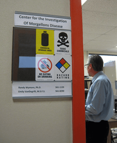
CENTER FOR INVESTIGATION OF MORGELLONS OKLAHOMA STATE UNIVERSITY-CENTER FOR HEALTH SCIENCES RANDY S. WYMORE, PH.D AUGUST 15, 2011

This summer the Center for the Investigation of Morgellons Disease (CIMD) at the Oklahoma State University Center for Health Sciences had an additional researcher. A summer scholar spent time in the lab complements of the Tulsa Area Biology Education Research Consortium.

The student spent over 160 hours doing microscopy and molecular biology under the supervision of Emily VanDegrift, MSFS. Research has focused on the microbiology and molecular biology of material associated with Morgellons Disease.

Approximately 40 solid and liquid media cultures were inoculated and grown under various conditions. Some media was fungal specific while others were suitable for growing various types of bacteria.

Individual colonies were grown from blood agar or standard lab media and the DNA extracted. The DNA was then amplified by PCR and analyzed. Some samples were purified and sent for DNA sequencing. DNA primers sensitive to all varieties of Pseudomonas & Cryptococcus produced no DNA when PCR amplification was performed.

While no PCR product was amplified from Morgellons samples the Pseudomonas & Cryptococcus positive controls all worked. This PCR product was examined on the highly sensitive genetic analyzer and suggests that, in the Morgellons samples and bacterial cultures grown, neither Pseudomonas nor Cryptococcus varieties were present in the Morgellons samples in question.

Thus far the DNA has most closely matched bacteria found typically in the gastro-intestinal tract and common skin bacteria. Samples are in the process of re-sequencing with different purification methods to more perfectly identify the specific strains, and to determine if any of the sequences have enough variability to suggest previously unknown bacterial types.

Typically this involves sequencing both strands of the DNA to eliminate any sequencing errors. No pathogenic or previously unknown strains have been observed thus far. Over the summer 30 sample collections have arrived from 4 countries and 3 continents, and are in the process of being examined. Pathology lab slides were submitted for evaluation with pathologists/histologists associated with OSU.

The biopsy slides revealed moderate inflammation with some infiltration by immune system cells. No fibers or other Morgellons-associated material were observed on the submitted slides.

Randy S. Wymore, Ph.D.
Director, OSU-CHS Center for the Investigation of Morgellons Disease
Associate Professor of Pharmacology
Oklahoma State University
Center for Health Sciences
1111 W. 17th St.
Tulsa, OK 74107

/wp-content/uploads/2020/09/CEHFLogo.png 0 0 Charles E. Holman Foundation /wp-content/uploads/2020/09/CEHFLogo.png Charles E. Holman Foundation2020-10-06 12:58:182021-01-12 15:51:21Research Update August 2011

Morgellons Study Cited by Faculty of 1000

Study of Emerging Skin Disease Among Top 2% Published

AUSTIN, Texas (June 19, 2012). A recent study of Morgellons disease has been cited as a “must read” by the Faculty of 1000 (F1000). The article entitled “Morgellons Disease: A Chemical and Light Microscopic Study”, by MJ Middelveen, EH Rasmussen, DG Kahn and RB Stricker, was published in the open‐access online Journal of Clinical and Experimental Dermatology Research. Three of the authors, including the principal investigator, serve without compensation on the Advisory Boards of The Charles E. Holman Foundation.

F1000 is a global community of over 10,000 experts who select, rate and evaluate the very best articles in biology and medicine. The service is widely used to find significant new research articles, and the inclusion of the Morgellons article places the work in the top 2% of published articles in these fields. The study evaluation can be accessed at the F1000 website ( http://f1000.com/716597867).In 2011, veterinary microbiologist Marianne J. Middelveen from Calgary, Alberta, Canada and internist Raphael B. Stricker, MD published a study documenting similarities between Morgellons disease and a veterinary illness known as bovine digital dermatitis (BDD) that causes lameness, decreased milk production, weight loss, and skin lesions near the hooves of affected cattle. That study revealed that the unusual fibers seen in the animal disease were similar to those seen in and under the skin of people worldwide who suffer from Morgellons disease. The new study confirms that Morgellons disease is not a delusional illness, as some in the medical community maintain.

The latest findings confirm that fibers from both bovine and human samples were similar in formation at the cellular level and had the chemical and physical properties of keratin. The keratin composition of filaments from humans was confirmed by immunohistological staining with antibodies specific for human keratins. Fibers from human patients were found to be biological in origin and are produced by keratinocytes in epithelial and follicular tissues. The findings are consistent with the 2012 Centers for Disease Control and Prevention (CDC) publication stating that over 80% of non‐biopsy material taken from patients had a protein composition.

“This study puts the final nail in the coffin of delusional disease that these patients have been labeled with,” stated Dr. Stricker. “It proves that Morgellons disease is a physiologic illness. From here on, scientists will be able to move forward in finding a cause and a cure.”

The Charles E. Holman Foundation is a 501(c) (3) nonprofit organization committed to advocacy and philanthropy in the fight against Morgellons disease. The foundation was named for Charles E. Holman, a pioneer in the fight against Morgellons disease. The Charles E. Holman Foundation is based in Austin, Texas and is led by Executive Director, Cindy Casey Holman, R.N.

For more information or to arrange interviews, please contact Ms. Cindy Casey, R.N.

/wp-content/uploads/2020/09/CEHFLogo.png 0 0 Charles E. Holman Foundation /wp-content/uploads/2020/09/CEHFLogo.png Charles E. Holman Foundation2020-10-06 12:53:282021-01-12 15:51:21Morgellons Study Cited by Faculty of 1000

OSU biomedical researcher says science, not belief, will prove existence of skin disorder Morgellons

/wp-content/uploads/2020/09/CEHFLogo.png 0 0 Charles E. Holman Foundation /wp-content/uploads/2020/09/CEHFLogo.png Charles E. Holman Foundation2020-10-06 12:44:342021-01-12 15:51:21OSU biomedical researcher says science, not belief, will prove existence of skin disorder Morgellons

LATEST RESEARCH

See the latest peer reviewed research publications >

CEHF on Blog-Talk Radio

The 6th Annual Medical-Scientific Conference on Morgellons

LISTEN NOW >

CEHMDF Refutes the CDC Report on Morgellons Disease

Featuring Presentations by:Peter Mayne and Randy Wymore

LEARN MORE >

Morgellons Study Cited by Faculty of 1000

Study of Emerging Skin Disease Among Top 2% Published

READ MORE >

The Charles E. Holman Foundation

PO Box 1109
Lone Star, TX
75668

events@thecehf.org

Neither the Charles E. Holman Foundation nor the individuals associated with it make any warranties or guarantees about any medical advice or treatment suggestions posted on this web site. This includes no guarantees about treatment effectiveness, long term or short term outcome, safety, or potential side effects. The choice to use any treatments posted on the web site is strictly left to the individual. The CEHF officially recommends you consult with your health care professional before using any medication or treatment, as only you and your medical care provider know your own unique and individual medical issues. While all treatment suggestions posted are reviewed by licensed medical personnel associated with the CEHF, the decision by an individual to use the information posted in their own treatment does not constitute the establishment of a doctor-patient relationship nor a contractual or professional arrangement of any sort.

© Copyright – Charles E. Holman Foundation

  • Home
  • Who We Are
  • What is Morgellons
  • Events
  • Contact Us
  • Donate

Scroll to top

We use cookies to enhance your browsing experience and analyze our traffic. By clicking "Accept All", you consent to our use of cookies.

Accept AllReject AllLearn More

Cookie and Privacy Settings


How we use cookies

We may request cookies to be set on your device. We use cookies to let us know when you visit our websites, how you interact with us, to enrich your user experience, and to customize your relationship with our website.

Click on the different category headings to find out more. You can also change some of your preferences. Note that blocking some types of cookies may impact your experience on our websites and the services we are able to offer.

Essential Website Cookies

These cookies are strictly necessary to provide you with services available through our website and to use some of its features.

Because these cookies are strictly necessary to deliver the website, refusing them will have impact how our site functions. You always can block or delete cookies by changing your browser settings and force blocking all cookies on this website. But this will always prompt you to accept/refuse cookies when revisiting our site.

We fully respect if you want to refuse cookies but to avoid asking you again and again kindly allow us to store a cookie for that. You are free to opt out any time or opt in for other cookies to get a better experience. If you refuse cookies we will remove all set cookies in our domain.

We provide you with a list of stored cookies on your computer in our domain so you can check what we stored. Due to security reasons we are not able to show or modify cookies from other domains. You can check these in your browser security settings.

Google Analytics Cookies

These cookies collect information that is used either in aggregate form to help us understand how our website is being used or how effective our marketing campaigns are, or to help us customize our website and application for you in order to enhance your experience.

If you do not want that we track your visit to our site you can disable tracking in your browser here:

Other external services

We also use different external services like Google Webfonts, Google Maps, and external Video providers. Since these providers may collect personal data like your IP address we allow you to block them here. Please be aware that this might heavily reduce the functionality and appearance of our site. Changes will take effect once you reload the page.

Google Webfont Settings:

Google Map Settings:

Google reCaptcha Settings:

Vimeo and Youtube video embeds:

Other cookies

The following cookies are also needed - You can choose if you want to allow them:

Privacy Policy

You can read about our cookies and privacy settings in detail on our Privacy Policy Page.

Accept settingsHide notification only