

Clinical Diagnosis of Morgellons Disease
Medical professionals often misdiagnose Morgellons disease symptoms. They may miscategorize them as other illnesses ranging from skin conditions to psychological disorders.
This leaves many patients without the care and treatments they need for relief.
All too frequently, doctors say nothing is physically wrong with their Morgellons patients. Instead, they attribute real symptoms to delusions of infestation—meaning the patient just thinks something has infected them.
However, evidence supporting an infectious cause is stacking up.
The majority of the medical community refuses to acknowledge Morgellons as an actual infectious disease, despite evidence on the contrary.
To develop a clinical diagnosis, a research team led by Marianne J. Middelveen has proposed criteria for identifying and classifying Morgellons disease.
Morgellons overview
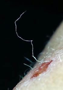
Morgellons is a complex skin disease characterized by ulcerating lesions (open sores) with protruding or embedded filaments.
These filaments or fibers are made from the patient’s skin cells that have gone hay-wire. They’re made of the same protein, keratin, and collagen that make our hair, skin, and nails.
Using hand-held light microscopes at 50x magnification, researchers can see filaments or fibers in the skin of Morgellons patients. The fibers may be blue, red, green, black, or white and are found growing under unbroken skin, protruding from calluses, and sprouting around skin lesions or open sores.
Aside from the strange fibers and obviously painful open sores, patients suffering from Morgellons disease also experience symptoms consistent with unresolved Lyme disease. These symptoms involve many different body systems and severely impact a patient’s quality of life. A patient with Morgellons may experience all of these symptoms:
- Extreme fatigue
- Muscle pain
- Arthritis
- Cardiac disease
- Memory loss
- Brain fog
- Depression and anxiety
- Disfiguring open sores
Morgellons Disease Fiber Extrusion
Through dozens of studies, researchers have discovered that 98% of Morgellons patients have an underlying Borrelia burgdorferi infection. Borrelia burgdorferi is a spirochetal bacteria spread through black-legged tick bites. They’re also the leading infectious agent in Lyme disease.
These spirochetes have been positively identified in patient tissue and body fluid samples using cell culture growths, PCR testing, SEM, TEM, light microscopy, and various staining techniques.
Morgellons patients account for up to 6% of Lyme disease diagnoses in Australia. This fact hints to researchers that persistent Lyme disease may manifest as Morgellons disease instead of a standalone illness.
The spirochetes in Lyme disease and Morgellons persists after antibiotic therapy through various adaptations that allow it to outrun the patient’s immune system. The little parasites hide inside specific cells and slow growth until the treatment stops.
Because of spirochete evasiveness, Morgellons disease is challenging to treat. Researchers and doctors haven’t yet discovered a practical course of antibiotic treatment that can permanently resolve the infection.
Clinical Diagnosis for Morgellons
Partly because of the treatment and diagnosis issues associated with Morgellons, Middelveen’s research team created a clinical diagnosis scheme for Morgellons. The idea behind the research is to help medical professionals accurately diagnose and evaluate the severity of their patients’ illnesses.
Medical classification schemes for disease staging help doctors objectively identify and assess disease severity and progression to prescribe the correct therapies and treatment
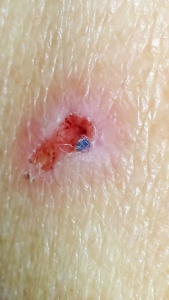
Morgellons Disease Lesion Close Up
routes. Syphilis, another spirochetal infection characterized by skin lesions, is categorized by three stages that reflect an array of skin sores and lesions associated with the disease.
Borrelia burgdorferi bacteria are the primary infectious agent that causes Morgellons disease. These flagellated spirochetes can “swim” through dense tissues and complex cellular structures, allowing them to travel or disseminate throughout the body of their hosts and infect a wide range of body tissues in various locations. In patients with a localized classification, the bacteria often haven’t yet spread beyond the body part that was initially bitten by the infectious tick.
The “class” designations in the staging for Morgellons disease denote how long the patient has been experiencing symptoms and whether the symptoms are local to one area of the body or disseminated to include others.
The “class” designations for the disease spread are paired with one of three varying stages of lesion progression. These stages help medical professionals assess the severity of the Borrelia infection. Stage A lesions are milder than Stage C lesions; Stage B lesions are somewhere between.
Below we share the specific classes and stages of Morgellons disease.
Class I
Early localized: lesions and fibers have been present for less than three months and are found on one central body area such as the head, trunk, or extremities.
Class II
Early disseminated: lesions and fibers have been present for less than three months and involve more than one area of the body.
Class III
Late, localized: fibers/lesions have been present for more than six months and are found on one area of the body, such as the head, trunk, or extremities.
Class IV
Late, disseminated: lesions and fibers have been present for more than six months and have spread to more than one body part. Patients may have lesions and fibers on their head, trunk, and extremities.
Stage A
During the first or early stage of Morgellons disease, the lesions have little immune cell reaction to the spirochetal infection. There are no macrophages (white blood cells) or hemorrhaging (bruising) in or around the lesions.
The skin cell layers are still organized, and the keratinocytes—cells that make keratin—have not been pushed into overdrive by the invading spirochetes.
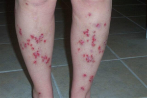
Painful lesions on a Morgellons patient’s legs
Stage B
Patients with Stage B lesions have an intermediary pattern that has progressed beyond Stage A but hasn’t yet reached the advanced degree seen in Stage C.
Stage C
White blood cells, macrophages, and Borrelia species colonies are easily found in late Stage C lesions samples. There’s remarkable bruising or hemorrhaging around the lesions, and the invading spirochetes have activated both Keratinocytes (keratin-producing cells) and fibroblasts (collagen-producing cells). The cells work in overdrive to produce unusual fibers and filaments. The skin cell layers in Stage C lesions have also been disrupted and are unorganized.
While identifying a clinical diagnosis can go a long way toward helping patients suffering from this disfiguring illness, there’s still much we don’t yet understand.
Donate today
Our researchers have made huge strides over the last decade to understand this debilitating illness better. But we are still far from reaching a cure or gaining traction in the mainstream medical community.
Only through grass-roots efforts can we spread awareness about the genuine impacts this disease has on Morgellons patients and their families.
Please do
nate today to the Charles E. Holman Morgellons Disease Foundation so we can continue to fund more research like this and spread awareness.